
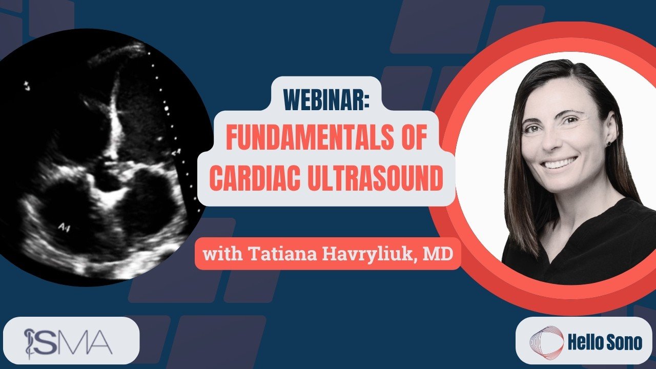
Fundamentals of Cardiac Ultrasound: Recognizing Critical Findings in Everyday Practice (Webinar Recording)
Read the related article on Hello Sono Blog at this link. You can view the full episode on YouTube at the link here.
Chest pain, dyspnea, hypotension, and suspected heart failure are among the most common reasons clinicians perform bedside cardiac evaluation.
Many providers still rely primarily on formal echocardiography and other imaging studies. But cardiac point-of-care ultrasound (POCUS) can rapidly provide critical bedside information about cardiac function, pericardial effusion, right heart strain, fluid responsiveness, and ejection fraction.
In this webinar, delivered in collaboration with the Southern Medical Association (SMA), we review the core cardiac ultrasound views, practical scanning techniques, and how cardiac POCUS can help guide real-time clinical decision-making in everyday clinical practice.
Watch the full recording:
What You’ll Learn About Cardiac Ultrasound
This practical, case-based session focuses on how to use cardiac POCUS to answer critical bedside questions in real time.
You’ll learn:
When cardiac POCUS is most helpful in patients with chest pain, dyspnea, hypotension, cardiac arrest, and suspected heart failure
How to obtain the core cardiac views, including parasternal long axis, parasternal short axis, apical four-chamber, subxiphoid, and IVC views
How to assess for pericardial effusion and cardiac tamponade
How to estimate ejection fraction using simple visual techniques
How to identify right heart strain and findings suggestive of pulmonary embolism
How IVC assessment helps evaluate fluid responsiveness and volume status
How cardiac POCUS findings guide rapid clinical decision-making in everyday clinical practice
Key Cardiac Ultrasound Findings Explained
Understanding core cardiac ultrasound findings is essential for bedside interpretation:
Pericardial Effusion: Anechoic fluid between the myocardium and pericardium
Cardiac Tamponade: Pericardial effusion with right atrial or right ventricular collapse and plethoric IVC
Right Heart Strain: Enlarged right ventricle, septal flattening (“D-sign”), and McConnell sign suggestive of pulmonary embolism
Reduced Ejection Fraction: Poor LV contractility, limited myocardial thickening, and increased EPSS
Hyperdynamic LV: Vigorous LV contraction that may suggest hypovolemia
Plethoric IVC: Dilated IVC with minimal respiratory collapse indicating elevated right-sided pressures
Collapsible IVC: Significant respiratory variation suggesting fluid responsiveness
Recognizing these patterns allows clinicians to answer focused clinical questions at the bedside.
Clinical Cases Reviewed
This webinar includes real-world cardiac POCUS cases demonstrating:
Pulmonary embolism with RV strain and D-sign
Cardiac tamponade with RV collapse
CHF exacerbation with poor ejection fraction and B-lines
Pericarditis with small pericardial effusion
Hyperdynamic LV in volume-depleted patients
Each case highlights how cardiac POCUS findings directly influence patient management.
Key Takeaways
Cardiac POCUS is a rapid bedside tool that helps answer focused clinical questions in real time
Simple cardiac views can provide critical information about cardiac function and hemodynamics
Estimating ejection fraction, evaluating for pericardial effusion, and identifying right heart strain are foundational skills
IVC assessment is most useful in patients with extreme volume findings
Clinical context should always be combined with cardiac, lung, and other POCUS findings
Why Clinicians Are Adopting Cardiac Ultrasound
Cardiac POCUS is increasingly used in urgent care, hospital medicine, and primary care because it can:
Accelerate bedside diagnosis
Improve hemodynamic assessment
Reduce delays in care
Support real-time treatment decisions
For many clinicians, cardiac POCUS becomes one of the most impactful bedside ultrasound applications in daily practice.
Next Steps: Building Confidence with POCUS
Learning cardiac ultrasound is the first step. Confidence comes from consistent use and feedback.
Hello Sono supports clinicians through:
Structured POCUS training
Longitudinal exam review and feedback
Implementation support
Learn more about the POCUS Exam Review: Here
At Hello Sono, we build high-quality, compliant, and financially sound POCUS programs to improve patient care and efficiency. Access the POCUS ROI Calculators to see the financial impact of POCUS. Fill out the contact form to speak to an expert.
Make sure to check out the Southern Medical Association page for more educational content.