
This case was originally published in the Journal of Urgent Care Medicine. The full article and accompanying discussion can be found here.
Case Overview
A 64-year-old woman came to urgent care with sudden, sharp pain in the back of her right ankle after stumbling on a curb that day.
She had trouble walking and climbing stairs.
For about 10 days before this, she had mild posterior ankle pain that she treated with ice, a compression wrap and crutches.
She had taken levofloxacin for a urinary tract infection about 2 weeks earlier.
Her history included diabetes, obesity, COPD and osteoarthritis.
Exam:
Bruising over the back of the ankle, spreading toward the lateral foot
Tenderness over the distal calf
Unable to actively plantarflex or do a single-leg heel raise
Thompson test inconclusive
POCUS Findings
A high-frequency linear probe was used over the posterior ankle.
The tendon ends were retracted, with fluid filling the gap between them.
This is consistent with a complete Achilles tendon rupture.
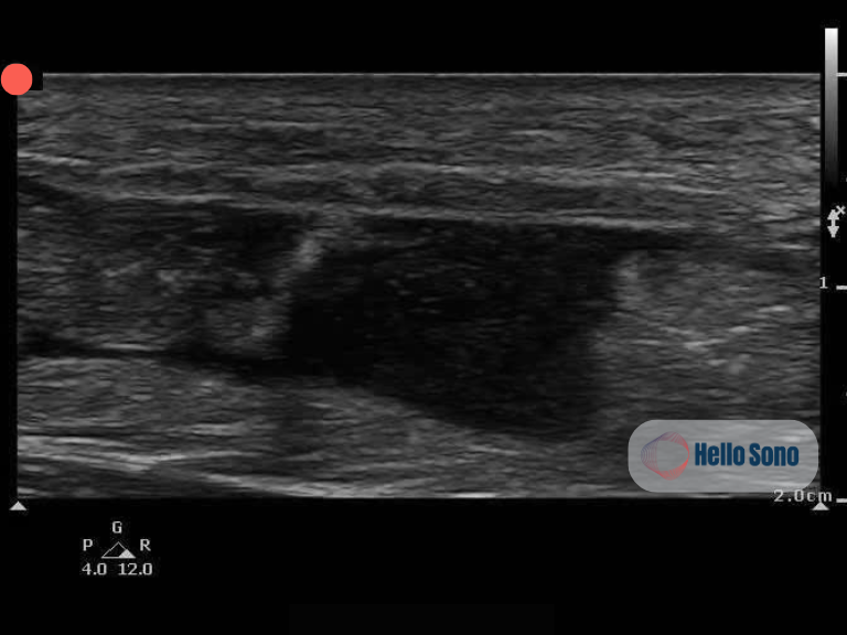
Ultrasound image of the Achilles tendon in the longitudinal plane shows the retracted torn tendon ends with a hypoechoic fluid-filled gap between them, consistent with with a complete Achilles tendon rupture.
Normal vs. Ruptured Tendon on Ultrasound
Normal: a continuous, bright, fibrillar band running from the myotendinous junction to the calcaneus.
Complete rupture:
Loss of fibrillar continuity
Retracted tendon ends
A fluid-filled gap between the ends signifying blood and inflammatory fluid
Scanning tip: scan the whole tendon in both long and short axis, from the myotendinous junction down to the calcaneal insertion.
Why This Case Matters
Achilles ruptures are often misdiagnosed as an ankle sprain or calf strain. This is especially likely when pain partly improves, or when the patient can still weakly plantarflex using accessory muscles.
A tendon already weakened by tendinopathy can make the presentation more subtle.
An equivocal Thompson test does not rule out rupture.
Risk factors to ask about:
Fluoroquinolones
Steroids
Oral bisphosphonates
Obesity
Diabetes
Chronic kidney disease
Hyperparathyroidism
Male sex
Weekend-warrior activity in deconditioned patients
Evidence
In a meta-analysis, ultrasound detected complete Achilles ruptures with a reported sensitivity of about 95% and specificity of about 99% (Aminlari et al., 2021). That study looked at surgically treated patients, so results may differ in a general urgent care population.
Partial tears are harder to see. Comparing with the other side and using dynamic scanning, such as a sonographic Thompson test, can help.
Pitfall: Don’t Mistake Bursitis for a Tear
Fluid in the retrocalcaneal bursa can look like a fluid-filled gap.
Always confirm whether the tendon fibers themselves are disrupted.
Evaluate for anisotropy (false positive) by rocking the probe. Learn more about anisotropy on our blog.
Impact on the Patient
Bedside ultrasound confirmed the diagnosis even though the exam was equivocal.
That allowed treatment to start right away:
Splinting in plantarflexion
No weight bearing
Urgent orthopedic referral
Timely recognition may help avoid a missed or delayed diagnosis.
Key Takeaway
Consider Achilles rupture in any patient with posterior ankle pain or bruising who can’t do a single-leg heel raise, even if the Thompson test is inconclusive. A focused POCUS exam of the full tendon can help confirm the diagnosis at the bedside. It works alongside clinical judgment and appropriate follow-up imaging, not in place of them.
For practices considering implementing or expanding POCUS programs, tools such as POCUS ROI Calculators and a POCUS Billing Readiness Checklist can help estimate financial impact and ensure appropriate workflow and documentation processes are in place.
References:
Reference
Aminlari A, Stone J, McKee R, et al. Diagnosing Achilles Tendon Rupture with Ultrasound in Patients Treated Surgically: A Systematic Review and Meta-Analysis. J Emerg Med. 2021;61(5):558-567.
Xergia SA, Tsarbou C, Liveris NI, et al. Risk factors for Achilles tendon rupture: an updated systematic review. Phys Sportsmed. 2023;51(6):506-516.
Fenech M, Ajjikuttira A, Edwards H. Ultrasound assessment of acute Achilles tendon rupture and measurement of the tendon gap. Australas J Ultrasound Med. 2024;27(2):106-119.
Griffin MJ, Olson K, Heckmann N, Charlton TP. Realtime Achilles Ultrasound Thompson Test for the Evaluation and Diagnosis of Acute Achilles Tendon Ruptures. Foot Ankle Int. 2017;38(1):36-40.