
Case Presentation:
A 38-year-old male presents to the Emergency Department with anterior knee pain after stumbling off a curb two days ago. He reports mild swelling above the kneecap but has full active range of motion and can perform a straight leg raise without difficulty. He denies any instability, locking, or prior knee injuries. Vital signs are normal. On exam, there is mild tenderness over the suprapatellar region without a palpable defect. The provider performs a point-of-care ultrasound of the quadriceps tendon. A longitudinal view of the distal quadriceps tendon is obtained with the knee in slight flexion. A focal hypoechoic region is noted within the quadriceps tendon.
What do you see, and what’s the diagnosis?
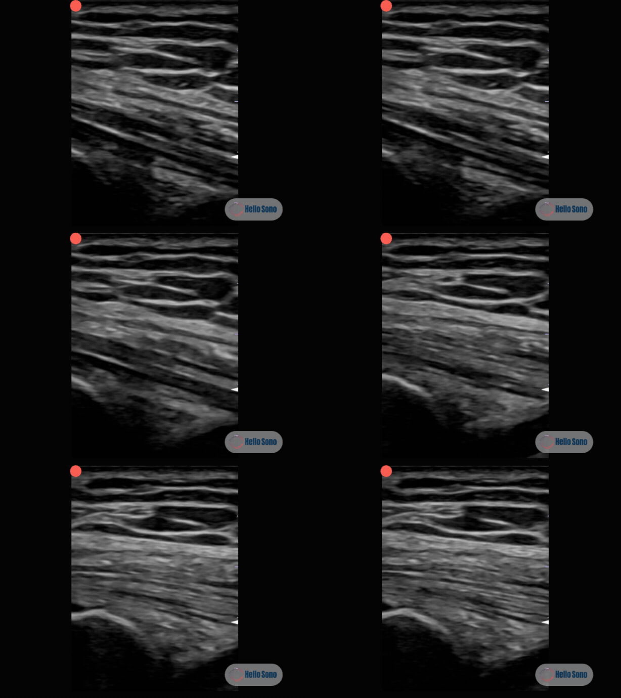
Figure 1: Longitudinal view of the distal quadriceps tendon showing a focal hypoechoic region within the tendon substance.
POCUS Findings:
The ultrasound demonstrates a focal hypoechoic region within the distal quadriceps tendon on longitudinal view, characterized by:
A dark zone located where the tendon curves toward the superior pole of the patella
Preserved fibrillar echotexture: the tendon fibers are not disrupted through the hypoechoic area
No anechoic gap, fluid collection, or tendon retraction
Complete resolution of the hypoechoic area when the probe is toggled to achieve perpendicular beam alignment
These findings are diagnostic of anisotropy artifact. In MSK ultrasound, anisotropy describes the way highly organized structures, especially tendons, can look falsely hypoechoic when the ultrasound beam is not perpendicular to the fibers. [3] The quadriceps tendon curves as it inserts on the patella, and when the ultrasound beam strikes the fibers obliquely, fewer sound waves reflect back to the transducer, creating a false hypoechoic region that can mimic a partial tear. In an ex vivo study, tendon echogenicity was shown to be highly angle-dependent: tendons become isoechoic to muscle at beam angles of just 2–7° off perpendicular and frankly hypoechoic at greater angles. [4] More recently, quantitative assessment of backscatter anisotropy has confirmed this in vivo, showing that normal patellar tendons exhibit significantly different anisotropy compared to tendons with tendinopathy, suggesting that changes in angle-dependent echogenicity can also be used to detect true tendon abnormalities. [7]
What is anisotropy and why is the quadriceps tendon especially vulnerable?:
Tendons are composed of highly organized, parallel collagen fibers. When the ultrasound beam is perpendicular (90°) to these fibers, sound waves reflect strongly back to the transducer, producing the characteristic bright, fibrillar echotexture. Even a small deviation from perpendicular causes a significant drop in echogenicity. [8] This is not a flaw in the ultrasound machine, it is a fundamental property of how organized collagen interacts with sound waves.
The quadriceps tendon is one of the most common sites for anisotropy artifact because:
It curves. The tendon follows the contour of the distal femur and curves anteriorly as it inserts on the superior pole of the patella. A flat probe cannot maintain perpendicular alignment across the entire tendon length simultaneously.
It is multi-layered. The quadriceps tendon is formed by the convergence of four muscle tendons (rectus femoris, vastus intermedius, vastus lateralis, and vastus medialis), creating layers with slightly different fiber orientations. The interfaces between layers can accentuate anisotropy.
It is superficial. High-frequency linear probes used for superficial structures have narrow beam widths, making them more sensitive to small angle changes.
The Fix: Adjusting the Angle of Incidence with Rocking and Fanning (Heel-Toe):
The bedside correction for anisotropy is simple and takes seconds. Beam angulation, either by physically rocking or fanning the probe or by using electronic beam steering on machines that support it, allows the operator to overcome the potential pitfall of anisotropy in ultrasound assessment of peripheral tendons [4]:
Identify the hypoechoic zone on the longitudinal view.
Gently rock (heel-toe) or fan (side to side on the short axis) the probe.
Watch the dark area in real time. If it "lights up" and returns to normal hyperechoic fibrillar echotexture, it was anisotropy.
Systematically rock through the entire tendon length to ensure every segment is evaluated with perpendicular beam alignment.
If the hypoechoic or anechoic area persists at every probe angle, it represents true pathology.
Additional techniques to reduce anisotropy include slight knee flexion (20–30°) to straighten the tendon and reduce curvature, and scanning in both longitudinal and transverse planes for complementary views.
What a Normal POCUS Should Show?:
In a normal quadriceps tendon with optimal probe positioning:
The tendon appears as a hyperechoic, fibrillar (striated) structure
The multi-layered architecture is visible: contributions from the rectus femoris (superficial), vastus intermedius (deep), and vastus lateralis/medialis
The tendon inserts smoothly on the superior pole of the patella
No focal hypoechoic or anechoic defects are present
The suprapatellar recess deep to the tendon contains minimal or no fluid
Normal tendons have a hyperechoic fibrillar echotexture that darkens and shows pattern disruption only in pathologic conditions. [10] Just as a normal LV should look like a symmetric donut on parasternal short-axis, a normal tendon should look like an unbroken, bright, striated band from origin to insertion when the beam is perpendicular.
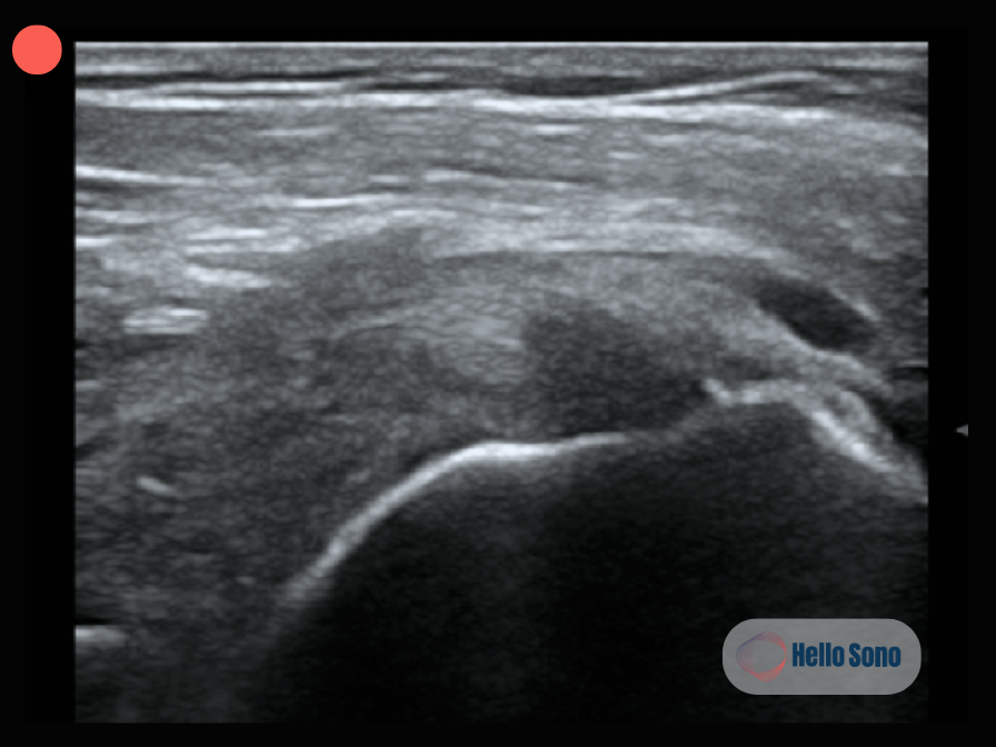
Figure 2. Longitudinal view of the supraspinatus tendon demonstrating a partial-thickness tear with a focal hypoechoic defect with disruption of the normal fibrillar pattern that persists regardless of probe angle, distinguishing true pathology from anisotropy artifact.
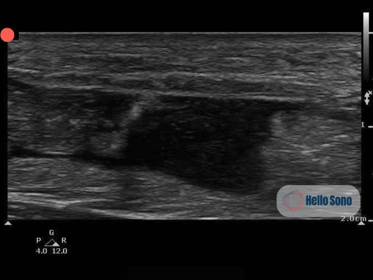
Figure 3. Longitudinal view of the Achilles tendon demonstrating a complete rupture with retraction of the proximal and distal tendon stumps and an anechoic fluid gap, representing the hallmark findings of a full-thickness tear.
Evidence:
Anisotropy is a well-established artifact in musculoskeletal ultrasound, first rigorously characterized in the late 1980s. An ex vivo study demonstrated that tendon echogenicity is highly angle-dependent, with tendons becoming isoechoic to muscle at beam angles of just 2–7° off perpendicular and hypoechoic at greater angles. [8] Beam angulation was subsequently described as a simple technique to overcome this pitfall in clinical practice. [4]
The clinical significance of anisotropy as a source of diagnostic error has been extensively documented in the shoulder. Anisotropy and transducer positioning have been identified as leading causes of false-positive sonographic findings of rotator cuff tears, alongside acoustic shadowing from the deltoid septum and anatomic variants at the rotator cuff interval. [3] Because the shoulder is primarily composed of nonlinear structures, the transducer should be continuously adjusted to maintain orthogonal clarity and prevent anisotropic artifact. [11]
For the quadriceps tendon specifically, ultrasound has been shown to be highly accurate for diagnosing tears when proper technique is used. A retrospective review of 239 consecutive knee sonograms reported 100% sensitivity, specificity, and accuracy for identifying high-grade partial tears and complete ruptures with surgical correlation. [5] An earlier surgical correlation study similarly reported 100% sensitivity and specificity for complete tears. [2] These high accuracy figures depend on the examiner's ability to distinguish true pathology from artifact, underscoring why anisotropy awareness is foundational to competent musculoskeletal POCUS.
In the emergency medicine setting, the 2023 ACEP Ultrasound Guidelines note that emergency physicians were able to diagnose extremity tendon injuries using POCUS with 100% sensitivity and 95% specificity in a prospective, multicenter study. [1,6] This supports the role of bedside ultrasound in acute tendon evaluation, provided the examiner is aware of artifacts like anisotropy that can lead to false-positive diagnoses.
More recently, quantitative assessment of backscatter anisotropy has been shown to differentiate normal tendons from those with tendinopathy, suggesting that the same angle-dependent property that causes diagnostic confusion may eventually be harnessed as a diagnostic tool. [7]
Case Resolution:
Given the patient's intact extensor mechanism, full active range of motion, and resolution of the hypoechoic zone with probe repositioning, the provider diagnoses anisotropy artifact of a normal quadriceps tendon. No tendon tear is present.
The patient is reassured, treated with ice, NSAIDs, and activity modification for his soft tissue contusion. He is discharged with return precautions for inability to straighten the knee, giving way, or worsening swelling. No advanced imaging is ordered.
Impact of POCUS:
In this case, recognizing anisotropy at the bedside:
Avoided a false-positive diagnosis of quadriceps tendon tear
Prevented unnecessary MRI and potential orthopedic referral
Reduced patient anxiety by providing immediate reassurance with a real-time demonstration, the provider was able to show the patient the dark zone disappearing as the probe was readjusted
Saved time and cost by resolving the clinical question in a single visit with no advanced imaging
Reinforced proper scanning technique, the fanning, rocking, and toggling maneuvers are teachable, reproducible skills
Understanding artifacts is just as important as recognizing pathology. A clinician who cannot identify anisotropy will overcall tendon tears; one who can will make confident, accurate diagnoses at the bedside. Emergency physicians can achieve 100% sensitivity and 95% specificity for tendon injuries with POCUS, but only when artifact recognition is part of the skill set. [1]
Conclusion:
Anisotropy is the most common artifact encountered in musculoskeletal POCUS and the leading cause of false-positive tendon "tears" at the bedside. The quadriceps tendon, with its curved insertion and multi-layered architecture, is particularly susceptible. The fix is simple: readjusting the angle of incidence of the probe until the beam is perpendicular to the tendon fibers. If the dark zone resolves, it was artifact. If it persists with fiber discontinuity, an anechoic gap, or dynamic separation, it is a real tear.
The same pitfall and the same fix apply across musculoskeletal POCUS: the rotator cuff, biceps tendon, Achilles tendon, and peripheral nerves are all vulnerable. Mastering the toggle maneuver is not an advanced skill, it is a foundational one.
Authored by Caleb Neal, DO, MPH
Reviewed by Tatiana Havryliuk, MD
Beyond clinical impact, integrating POCUS into clinical workflows enables appropriate reimbursement when supported by proper credentialing, documentation, and quality assurance. For a practical framework, download the Hello Sono POCUS Billing Readiness Checklist.
References:
ACEP Ultrasound Guidelines: Emergency, Point-of-Care, and Clinical Ultrasound Guidelines in Medicine. 2023.
Bianchi S, Zwass A, Abdelwahab IF, Banderali A. Diagnosis of Tears of the Quadriceps Tendon of the Knee: Value of Sonography. AJR Am J Roentgenol. 1994;162(5):1137-1140.
Connolly DJ, Berman L, McNally EG. The Use of Beam Angulation to Overcome Anisotropy When Viewing Human Tendon With High Frequency Linear Array Ultrasound. Br J Radiol. 2001;74(878):183-185.
Crass JR, van de Vegte GL, Harkavy LA. Tendon Echogenicity: Ex Vivo Study. Radiology. 1988;167(2):499-501.
Foley R, Fessell D, Yablon C, et al. Sonography of Traumatic Quadriceps Tendon Tears With Surgical Correlation. J Ultrasound Med. 2015;34(5):805-810.
Gao J, Rosander A, Rubin JM. Quantitative Assessment of Tendon Backscatter Anisotropy in B-Mode Ultrasound. Ultrasound Med Biol. 2023;49(6):1407-1413.
Jacobson JA. Shoulder US: anatomy, technique, and scanning pitfalls. Radiology. 2011 Jul;260(1):6-16. doi: 10.1148/radiol.11101082. PMID: 21697306.
Rutten MJ, Jager GJ, Blickman JG. US of the Rotator Cuff: Pitfalls, Limitations, and Artifacts. Radiographics. 2006;26(2):589-604.
Strakowski JA, Chiou-Tan FY. Musculoskeletal Ultrasound for Traumatic and Torsional Alterations. Muscle Nerve. 2020;62(6):683-697.
Strakowski JA, Visco CJ. Diagnostic and Therapeutic Musculoskeletal Ultrasound Applications of the Shoulder. Muscle Nerve. 2019;60(1):1-6.
Wu TS, Roque PJ, Green J, et al. Bedside Ultrasound Evaluation of Tendon Injuries. Am J Emerg Med. 2012;30(8):1357-1362.