
Gallbladder Ultrasound for Everyday Practice (Webinar Recording)
Read the related article on Hello Sono Blog at this link. You can view the full episode on YouTube at the link here.
Right upper quadrant (RUQ) pain, nausea, vomiting, and fever are common clinical presentations where point-of-care ultrasound (POCUS) can provide valuable bedside information.
While some patients require additional imaging or emergency evaluation, gallbladder POCUS can help clinicians rapidly assess for cholelithiasis, evaluate for signs of acute cholecystitis, and support clinical decision-making in the appropriate clinical context.
In this webinar, presented in collaboration with the Southern Medical Association (SMA), we review a practical approach to gallbladder ultrasound, including probe selection, patient positioning, image acquisition, and how POCUS findings can be integrated into the evaluation of patients with suspected gallbladder and biliary pathology.
Watch the full recording:
What You’ll Learn About Gallbladder Ultrasound
This practical session focuses on performing and interpreting gallbladder POCUS in everyday clinical practice.
You’ll learn:
Common indications for gallbladder POCUS
How to select the appropriate probe and position the patient to obtain diagnostic-quality long- and short-axis gallbladder views
How to obtain key measurements, including anterior gallbladder wall thickness and common bile duct (CBD) diameter
How to identify the portal triad using the "Mickey Mouse sign"
The sonographic appearance of cholelithiasis, gallbladder sludge, and gallbladder polyps
How to recognize acute cholecystitis
How to identify the WES (wall-echo-shadow) sign and recognize findings suggestive of choledocholithiasis
Common pitfalls, such as mistaking the duodenum for gallbladder pathology
How gallbladder POCUS findings can support clinical decision-making alongside the patient's history, physical examination, and other diagnostic information
Key GallBladder Ultrasound Findings Explained
Recognizing common ultrasound findings is essential for accurate bedside interpretation.
Normal Gallbladder: A thin-walled, anechoic, distended structure without stones, sludge, or pericholecystic fluid.
Cholelithiasis: Echogenic, mobile structures within the gallbladder lumen that produce posterior acoustic shadowing.
Gallbladder Sludge: Low-level, non-shadowing echogenic material layering within the gallbladder that moves slowly with patient repositioning.
Gallbladder Polyps: Echogenic structures projecting into the gallbladder lumen that do not shadow and remain fixed to the wall regardless of patient position.
Acute Cholecystitis: Suggested by a combination of findings, including pericholecystic fluid, anterior gallbladder wall thickness greater than 3 mm, and a positive sonographic Murphy sign.
WES (Wall-Echo-Shadow) Sign: A pattern seen when the gallbladder is contracted around stones, producing a characteristic sequence of wall, echo, and shadow that can obscure the underlying gallbladder.
Choledocholithiasis: Suggested by a dilated common bile duct, generally greater than 6 mm, and warrants further evaluation.
Mickey Mouse Sign: A cross-sectional view of the portal triad in which the portal vein and adjacent hepatic artery and common bile duct create an appearance resembling a familiar cartoon character, used to help identify and measure the CBD.
A common pitfall is mistaking a fluid-filled loop of duodenum adjacent to the gallbladder for gallbladder pathology; using peristalsis and probe manipulation can help distinguish bowel from the gallbladder.
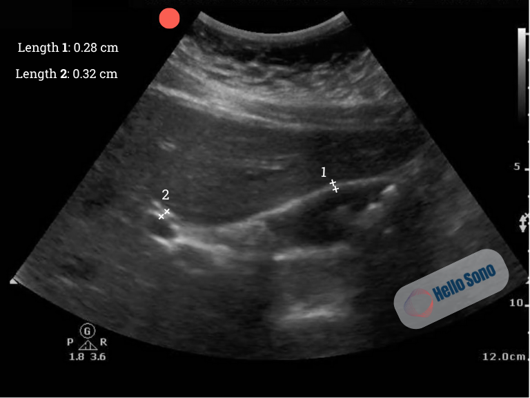
Ultrasound image of the gallbladder in long-axis demonstrating gallstones at the fundus and normal CBD and anterior gallbladder wall measurements.
Key Takeaways
1. Fast bedside answers
POCUS quickly shows gallstones, cholecystitis, or a dilated common bile duct in patients with RUQ/epigastric pain, nausea, fever, or jaundice.
2. Positioning matters
Left lateral decubitus positioning combined with a deep breath generally provides the clearest gallbladder view.
3. Know the cholecystitis findings
Pericholecystic fluid, stones or sludge, wall thickness over 3 mm, and a positive sonographic Murphy's sign are key findings to assess.
4. Watch for pitfalls
Avoid mistaking the duodenum for the gallbladder and remain alert for the WES sign.
Why Clinicians Are Incorporating Gallbladder POCUS
Gallbladder POCUS is increasingly used across emergency medicine, primary care, urgent care, and hospital medicine because it can:
Provide immediate bedside information during patient evaluation
Help identify cholelithiasis and sonographic signs of acute cholecystitis
Support clinical decision-making when evaluating suspected biliary pathology
Reduce unnecessary delays while determining the next diagnostic or management step
Complement the physical examination and other diagnostic testing
For many clinicians, gallbladder POCUS has become a valuable component of the bedside assessment of patients with suspected biliary tract pathology.
Next Steps: Building Confidence with POCUS
Learning gallbladder ultrasound is only the beginning. Developing confidence requires continued practice, image review, and feedback.
Hello Sono supports clinicians through:
Structured POCUS training
Longitudinal exam review and feedback
Implementation support
Learn more about the POCUS Exam Review: Here
At Hello Sono, we build high-quality, compliant, and financially sound POCUS programs to improve patient care and efficiency. Access the POCUS ROI Calculators to see the financial impact of POCUS. Fill out the contact form to speak to an expert.
Make sure to check out the Southern Medical Association page for more educational content.