
A Young Man with Chest Pain: When POCUS Makes the Difference
This article also appears in the Southern Medical Association News at this link.
Case Presentation:
A healthy 25-year-old male with no significant past medical history presented to a rural clinic with sudden-onset, sharp right-sided chest pain that began earlier in the day while walking. The pain was pleuritic, but he denied shortness of breath, fever, trauma, or recent illness. On physical exam, his vitals were normal, lung sounds were clear bilaterally, and there was no chest wall tenderness or crepitus. An EKG was performed and found to be normal. With no chest X-ray available onsite, the provider performed a point-of-care ultrasound (POCUS) of the lungs. The representative clip for the right lung apex is shown below.
What do you see, and what’s the diagnosis?
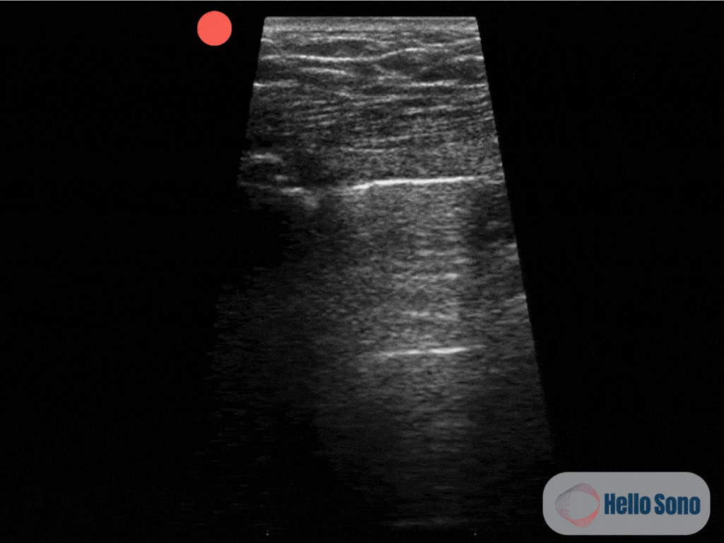
Clip 1: This ultrasound clip shows the apex of the right lung. The pleura appears as a bright white (hyperechoic) horizontal line near the top of the image. You can observe pleural sliding on the right side of the image, while the left side shows no sliding.
POCUS Findings:
The ultrasound clip reveals a lung point, the exact location where normal lung with pleural sliding transitions to an area without sliding. This “on-off” pattern is pathognomonic for pneumothorax. On one side of the image, shimmering pleural sliding confirms lung apposition; on the other side, the absence of sliding confirms the presence of air in the pleural space. See annotated clip (Clip 2).
Although complete absence of lung sliding may suggest a pneumothorax, it is not specific. Other conditions, such as pleural adhesions, large subpleural blebs, subpleural pneumonia, or scarring from prior infection or surgery, can also eliminate lung sliding. Therefore, identifying the lung point is key as it confirms the presence of a pneumothorax with high specificity.
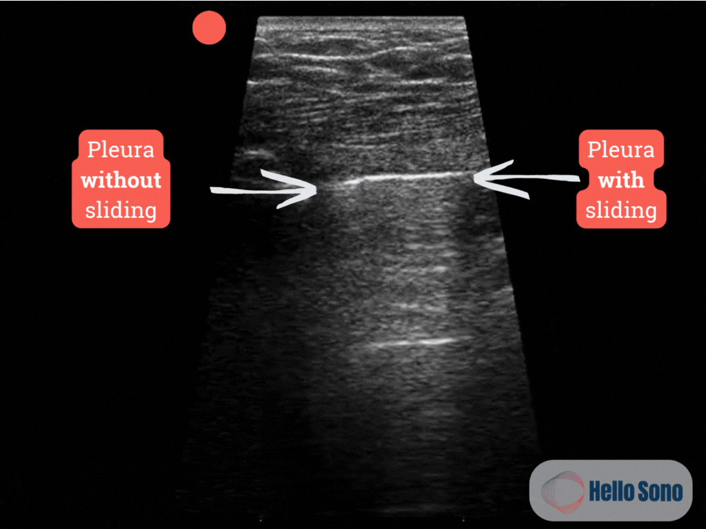
Clip 2: Ultrasound clip showing the lung point at the apex of the right lung with annotations.
In addition, the location of the lung point can help estimate the size of the pneumothorax. More anterior lung points are typically associated with smaller pneumothoraces, while more lateral or posterior lung points suggest a larger volume of air in the pleural space. In this case, the lung point was found at the apex of the right lung, suggesting that the pneumothorax is very small in size.
What a Normal POCUS Should Show:
In a healthy individual, lung POCUS shows continuous pleural sliding, a shimmering horizontal motion of the visceral and parietal pleura during respiration (Clip 3).
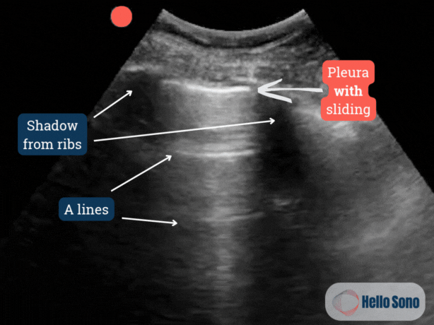
Clip 3: Ultrasound clip of the normal lung showing pleural sliding with annotations.
Evidence:
Lung ultrasound has been shown to outperform chest X-ray in detecting pneumothorax. For example, a 2020 Cochrane review found the sensitivity of lung ultrasound to be 91% (95% CI 85–94%) compared to 47% (95% CI 31–63%) for supine chest radiography in trauma patients. Similarly, the Society of Critical Care Medicine, in its guidelines, states that the sensitivity of ultrasound exceeds 85%, compared with approximately 30–75% for conventional radiography. [1,2] The specificity of the lung point sign on lung ultrasound for the diagnosis of pneumothorax is 100%. [2,3] Not only is POCUS more sensitive and specific, but it’s also faster, repeatable, and can be performed in settings where X-ray is delayed or unavailable, like rural or resource-limited clinics.
Case Resolution:
The identification of a lung point on POCUS confirmed the diagnosis of a right-sided pneumothorax. Judging by its location at the apex of the lung, this was a small pneumothorax. Although the patient was hemodynamically stable and asymptomatic apart from chest pain, he was referred to the nearest emergency department for monitoring and further evaluation. While outpatient management may eventually be an option in select cases, most rural clinics lack the resources to safely observe these patients, making ED referral the most appropriate choice.
Impact of POCUS:
In this rural clinic, POCUS enabled a rapid, definitive diagnosis of pneumothorax in a patient who might otherwise have experienced a significant delay. It allowed the provider to triage appropriately, initiating care that could prevent progression to a life-threatening tension pneumothorax. For rural clinicians, lung ultrasound is a critical, high-yield tool that can be quickly learned and integrated into everyday practice. Learn about other high-yield POCUS applications in primary and urgent care.
Conclusion:
Pneumothorax is a life-threatening condition that must be ruled out in patients presenting with pleuritic chest pain or undifferentiated dyspnea. In rural settings, where imaging access is often limited, POCUS offers a powerful alternative. The ability to identify a lung point provides a definitive diagnosis and supports timely and appropriate patient transfer or treatment. For more on lung ultrasound in the evaluation of dyspnea, see our prior article on differentiating CHF from COPD exacerbations using POCUS.
Ready to take the next step with POCUS? Hello Sono helps practices roll out high-quality, compliant, and profitable POCUS programs.
Authored by Dr. Tatiana Havryliuk
References:
Chan KK, Joo DA, McRae AD, Takwoingi Y, Premji ZA, Lang E, Wakai A. Chest ultrasonography versus supine chest radiography for diagnosis of pneumothorax in trauma patients in the emergency department. Cochrane Database Syst Rev. 2020 Jul 23;7(7):CD013031. doi: 10.1002/14651858.CD013031.pub2. PMID: 32702777; PMCID: PMC7390330.
Frankel HL, Kirkpatrick AW, Elbarbary M, et al. Guidelines for the Appropriate Use of Bedside General and Cardiac Ultrasonography in the Evaluation of Critically Ill Patients—Part I: General Ultrasonography. Critical Care Medicine 43(11):p 2479-2502, November 2015. | DOI: 10.1097/CCM.0000000000001216
Lichtenstein DA, Mezière G, Lascols N, Biderman P, Courret JP, Gepner A, Goldstein I, Tenoudji-Cohen M. Ultrasound diagnosis of occult pneumothorax. Crit Care Med. 2005 Jun;33(6):1231-8. doi: 10.1097/01.ccm.0000164542.86954.b4. PMID: 15942336.