
Renal & Bladder POCUS: A Practical Guide to Evaluating Kidney Stones & Urinary Retention (Webinar Recording)
Read the related article on Hello Sono Blog at this link. You can view the full episode on YouTube at the link here.
Flank pain, hematuria, urinary retention, and acute kidney injury (AKI) are common clinical presentations where point-of-care ultrasound (POCUS) can provide valuable bedside information.
While some patients require additional imaging or emergency evaluation, renal and bladder POCUS can help clinicians rapidly assess for hydronephrosis, evaluate bladder distention, and support clinical decision-making in the appropriate clinical context.
In this webinar, presented in collaboration with the Southern Medical Association (SMA), we review a practical approach to renal and bladder ultrasound, including scanning technique, image interpretation, and how POCUS findings can be integrated into the evaluation of patients with suspected renal colic and urinary retention.
Watch the full recording:

What You’ll Learn About Renal & Bladder Ultrasound
This practical session focuses on performing and interpreting renal and bladder POCUS in everyday clinical practice.
You’ll learn:
Common indications for renal and bladder POCUS, including suspected renal colic, urinary retention, hematuria, and AKI
How to obtain high-quality kidney and bladder views using appropriate probe positioning and scanning techniques
The sonographic appearance of normal renal anatomy and the collecting system
How to identify and grade hydronephrosis
How to distinguish hydronephrosis from common mimics such as renal cysts and normal renal vasculature
How bladder ultrasound can help assess post-void residual volume and urinary retention
How renal and bladder POCUS findings can support clinical decision-making alongside the patient's history, physical examination, and other diagnostic information
Key Renal & Bladder Ultrasound Findings Explained
Recognizing common ultrasound findings is essential for accurate bedside interpretation.
Normal Kidney: A normal collecting system appears collapsed and echogenic without dilation.
Hydronephrosis: Dilation of the renal collecting system that may be graded as mild, moderate, or severe based on the extent of collecting system dilation and cortical thinning.
Severe Hydronephrosis: Marked collecting system dilation accompanied by cortical thinning, suggesting prolonged obstruction and the need for emergent decompression if the finding is acute.
Simple Renal Cyst: A well-defined anechoic structure that does not communicate with the collecting system and typically does not require follow-up when it has benign sonographic features.
Complex Renal Cyst: A cyst with internal echoes, septations, or other complex features that warrants further evaluation.
Urinary Retention: An enlarged post-void bladder volume consistent with incomplete bladder emptying, which should be interpreted within the clinical context.
Using color Doppler when appropriate can help distinguish hydronephrosis from adjacent renal blood vessels.
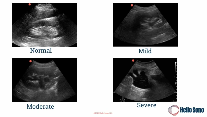
Understanding the sonographic appearance of normal, mild, moderate, and severe hydronephrosis is a foundational skill for clinicians performing renal point-of-care ultrasound (POCUS).
Key Takeaways
Renal and bladder POCUS provides rapid bedside assessment for hydronephrosis and bladder distention.
Hydronephrosis should be graded systematically, as the severity of findings may influence further evaluation and management.
Color Doppler can improve diagnostic confidence when differentiating hydronephrosis from renal vasculature.
Renal cysts and other sonographic mimics should be recognized to avoid misinterpretation.
POCUS findings should always be interpreted in conjunction with the patient's clinical presentation and used to guide, rather than replace, clinical judgment.
Why Clinicians Are Incorporating Renal & Bladder POCUS
Renal and bladder POCUS is increasingly used across emergency medicine, primary care, urgent care, and hospital medicine because it can:
Provide immediate bedside information during patient evaluation
Help identify hydronephrosis and urinary retention
Support clinical decision-making when evaluating suspected renal colic
Reduce unnecessary delays while determining the next diagnostic or management step
Complement the physical examination and other diagnostic testing
For many clinicians, renal and bladder POCUS has become a valuable component of the bedside assessment of patients with suspected urinary tract pathology.
Next Steps: Building Confidence with POCUS
Learning renal and bladder ultrasound is only the beginning. Developing confidence requires continued practice, image review, and feedback.
Hello Sono supports clinicians through:
Structured POCUS training
Longitudinal exam review and feedback
Implementation support
Learn more about the POCUS Exam Review: Here
At Hello Sono, we build high-quality, compliant, and financially sound POCUS programs to improve patient care and efficiency. Access the POCUS ROI Calculators to see the financial impact of POCUS. Fill out the contact form to speak to an expert.
Make sure to check out the Southern Medical Association page for more educational content.